RANGE OF SERVICESAngiography Outpatient Clinic |
||||||||||||
|
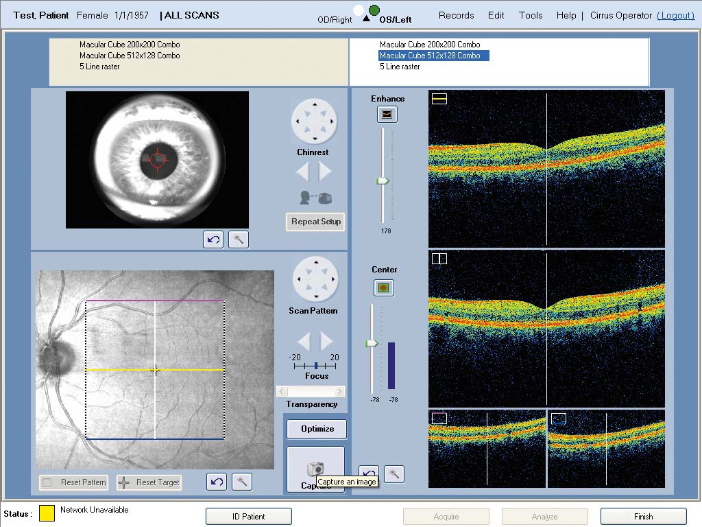
The Angiography Outpatient Clinic performs the following activities: Fluorescein angiography OCT - optical coherence tomography
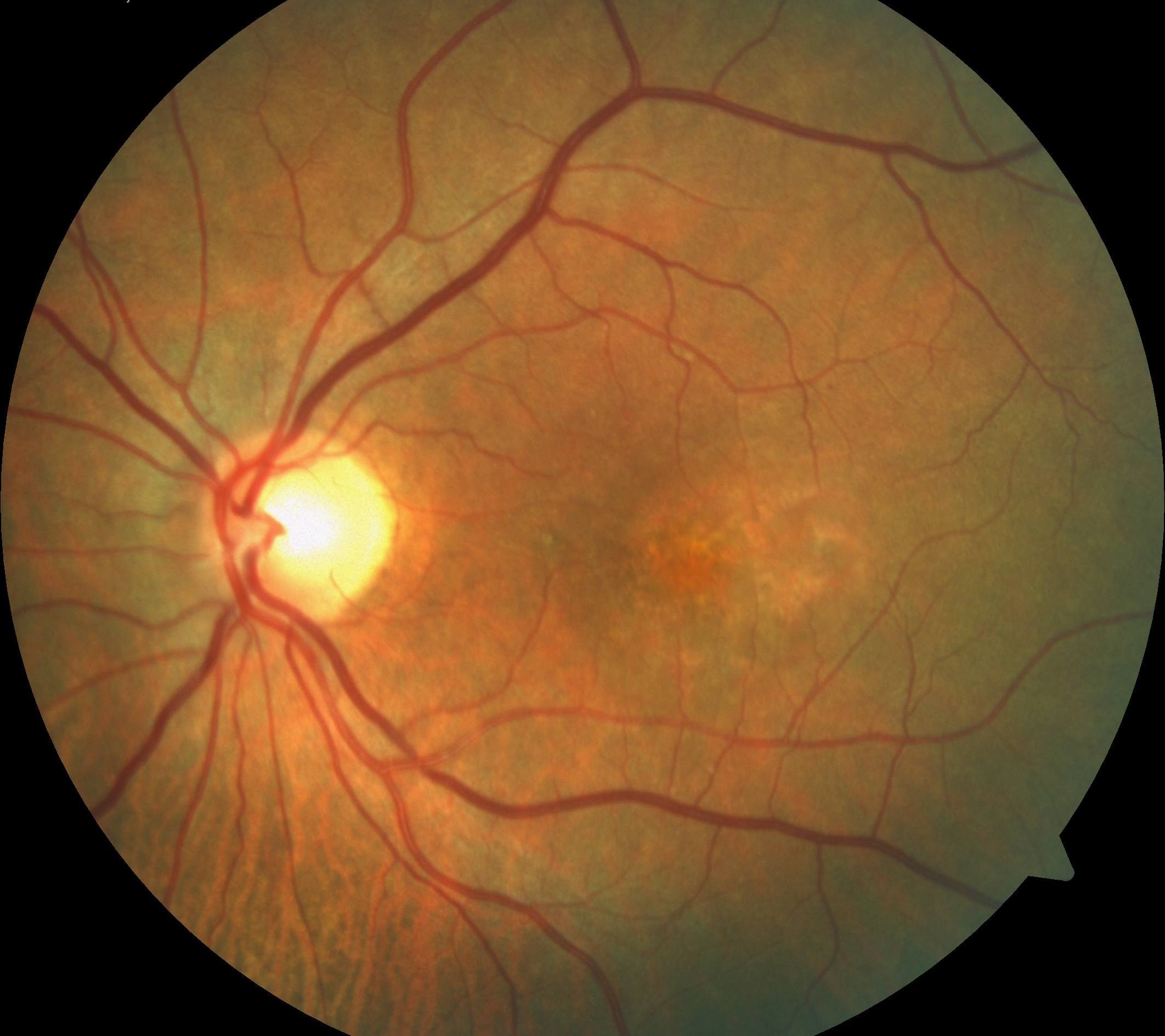
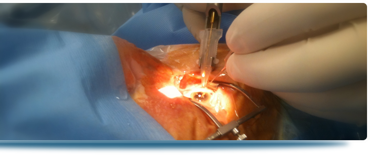
Fluorescein angiography The technique of fluorescein angiography was first described by Angus MacLean and A. Edward Maumenee nearly 50 years ago. In the recent years the technique has developed as the one widely used for examining the circulation of the retina and choroid. Angiography has become one of the basic diagnosis techniques in the following disorders: Diabetes and its complications – the test allows to qualify the patient to laser therapy (photocoagulation) AMD - Age-related Macular Degeneration – distinguishing its wet and dry type. Analysing angiography together with OCT enables to qualify to imply anti-VEGF medications. Eye cancers – the test allows to discriminate between cancers and inflammatory or degeneration condition. Angiography is a technique using a fluorescent dye and a specialized camera that takes photos of the eye fundus. The test begins with giving eye drops in order to cause dilation of the pupils and inserting a small intravenous cannula at the front of an elbow or at the back of a hand. Fluorescein – a type of dye - is given through the cannula and it reaches the eye blood vessels in a few seconds. Then digital photographs are taken. Angiography is a safe technique which rarely results in serious side effects. A small number of patients may feel sick. After the test one may see everything in red colour which is a temporary result of the camera strong flashlight. Allergic reactions and asthma that are the most serious side effects occur very rarely. In our clinic angiography is done under local anaesthesia.
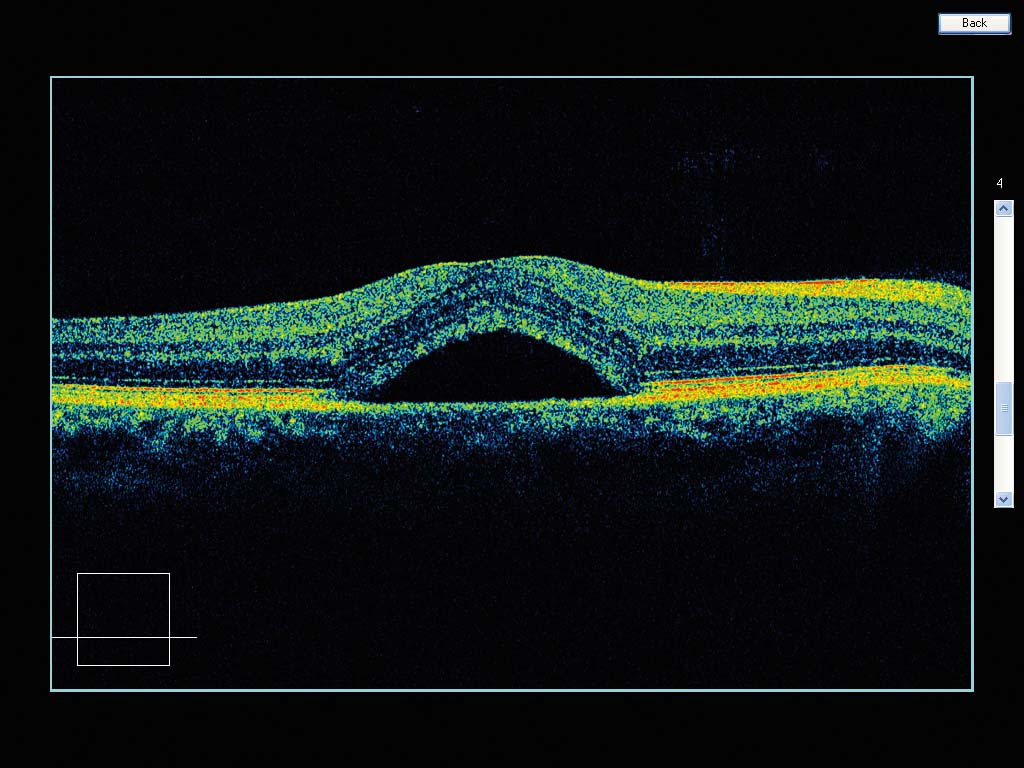
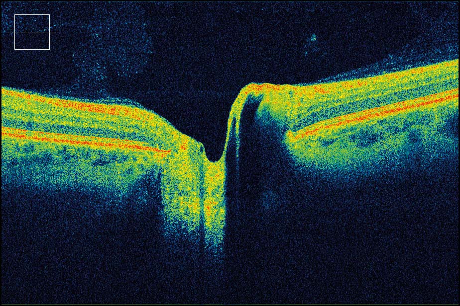
OCT - optical coherence tomography OCT is a modern method of imaging and diagnostics in eye diseases much more precise than magnetic resonance imaging (MRI), computer tomography or USG. It is used to anterior segment of eye and retina imaging. It is a completely non-invasive test – the device does not touch the eye and the dye is not used here. The device used in our center allows to get 5- micrometer-resolution images. OCT often complements fluorescein angiography or sometimes even replaces it. Optical coherence tomography enables to visualize the macula, the optic nerve, it allows to watch the retina layers and the open- or closed-angle glaucoma. The technique is essential in modern diagnostics and monitoring such diseases as glaucoma, AMD, retinal detachment, epiretinal membrane, macular edema or macular holes.
|
number of visitors: 939894 OCULUS Eye Diseases Treatment Center | 3 Feliksa Nowowiejskiego Street | 75-587 Koszalin
design: internet-media.pl
